Thursday 26 June 2008

exam question


click to enlarge.

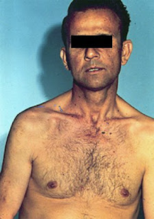
a friend of mine had his exam recently.
he had a tall patient in station 5 and was asked to come with a diagnosis.
he was confused between Marfan's and Klinefelter's syndromes!
how would you differentiate?

Infective Endocarditis


What are the update in NICE guidelines March 2008 regarding prophylaxis against infective endocarditis?

Wednesday 25 June 2008

Child-Pugh score


click to enlarge.
what do you know about Rockall scoring system?

important acts to remember

the mental Capacity act 2005:
this act provides a legal framwork for making decisions in relation to people who lack capacity.
link

the mental health act 2007:
this act provides a statutory framework, which sets out when patients can be compulsorily treated for mental disorder without consent,to protect them or others from harm.
link

the human tissue act 2004:
this act require consent to be obtained before a person's tissue can be stored and used.
link

the human fertilisation and emberyology act 1990:
this act privide legal framework across UK for all those involved in fertility treatment.
currently the parliament is considering number of amendments to the act.
link

the human right act 1998:
the right to life and the right to be free from degrading treatment.
link

Tuesday 17 June 2008

systolic murmurs

one of the common problems in the cardiology station is confusion regarding the systolic murmur.is is MR or AS?!! or may be VSD?
practicing is the only way.
here is a reminder:


click to enlarge


remember:
*practice,lestin to as many as you can.
*try to diagnose the valve lesion before you apply your stethoscope-attention to pulse character, BP loction of the apex.
*character of the murmur often more useful than its location.
*if you have no clue what is the diagnosis, describe your findings, you can still gain some points.

try to identify the systolic murmurs below!

systolic murmur 1

what is the valve lesion?

systolic murmur 2

what is the valvular lesion?

systolic murmur 3.

what is the valvular lesions?

check list...severity and management


click to enlarge

Saturday 14 June 2008

exam question 8


what is the most likely diagnosis?
how would you manage it?

check list...remember


click to enlarge

cerebellar syndrome


Examination of the cerebellar system:
Check eye movement for nystagmus
“..Put your arms straight in front of you...”
Intension tremor
Diminished/rebound reflexes
Examine for coordination
Finger-nose test
Rapid alt movements
Check for dys synergia
Check for coordination in the legs
Assess gait, Assess speech
Examine fundi for optic atrophy

what are the causes of cerebellar syndrome?
read more

Friday 13 June 2008

Hand examination

Inroduce yourself
Explain about your task
Ask about any pain
Correct positioning of the hands e.g pillow
•inspect the dorum for:
Nails pitting,vasculitis,atrophy
Skin
Small muscle wasting guttering
Deformity (memorise them)
•inspect the palmer aspect for:
Pulp top
Erythema
Wasting of the thener and hypothener
Contracture
Scar of decompansation
•Back to the dorsum:
Palpate the joint for evidance of synovitis
Palpate the wrist
•Back to the palmer aspect:
Perform tenil test
•check the motor system in the hand:
“squeeze my fingers” C8 T1
“hold your fingers striaght” stop me from pending them C7
“spread your fingers”stop me pushing them togerther ulnar
“point your thumb to ceiling”stop me pushing it down median
•check sensation
•Feel for subcut nodules
•Assess disability

Thursday 12 June 2008

cardiovascular examination


follow these steps in CVS examination:

Introduction : Hello Mr Smith,

My name is Dr X ; I'm a medical SHO,

Can I examine your heart please?

Position : 45 degree

Is it sore anywhere?

Exposure: can you take your shirt please?

General inspection : stand back,

Ask the patient to take deep breath in,

Look is he comfortable at rest, is he SOB?

Look for catheter, peripheral oedema, GTN, OXYGEN,

Malar rash,Lip cyanosis, BP apparatus.

Hands : look for warm/cold

Clubbing

Splinter haemorrhage

Xanthoma

Tobacco staining

Arm: pulse (rate, rhythm, volume, character & synchronous) Radio-radial delay

Collapsing pulse (ask for pain in shoulder)

Radio-femoral delay

Brachial pulse & anti cubital fosaa scar.

Feel carotid pulse & comment on character.

Face:

eyes for paller, xanthlasma

Mouth for tongue, cyanosis

JVP

Inspection:

Comment on mid sternal scar/ mitral valvotomy scar.

Palpation:

Palpate for apex beat (count the apex beat from sternal angle.

comment on position and character? tapping/heaving/normal)

Palpate for heave on either side of sternum

Palpate for thrill in 2 ICS

Auscultation:

Start from the apex. Comment on s1-s2

Always sync with carotid pulse and also ask pts to breath

in and out to look for respiratory variable

If PSM of MR then radiate to axilla

If EDM of MS (if u cant here murmur then L lateral position with bell)

PALPATE APEX BEFORE AUSCULTATE.

If ESM of AS auscultate in apex, below sternum,

left 2nd ICS, right 2nd ICS, then to carotid pulse.

Comment on site of best audible.

If EDM ofAR auscultate in right 2nd with pts leaning

forward with breath held in expiration.

when finished,

Thank the pt

Conclude by saying:

I would like to finish ex by

checking signs of heart failure

(auscultating lung bases, looking for hepatomegaly, pedal oedema and sacral oedema).

Checking BP.

Checking urine

the jugular venous pulse

examine this patient's neck.
comment on the waveforms.
how to time the waves of the JVP with the carotid pulse?

exam question


what is the name of this sign?
what are the features of the underlying condition?
if you have asked to lestin to this patients' heart,what murmur would you expect?

should score 4 in scar!


lestin to the heart of this patient.
what is your diagnosis?(two possibilities)
this patient present with bleeding.what is the most likely cause and how would you manage it?

scar in cardiovascular station

examples regarding consent and capacity

Examples from GMC

Consent: patients and doctors making decisions together.


Refusal of treatment:

C had paranoid schizophrenia and was detained in Broadmoor Secure hospital. He developed gangrene in his leg but refused to agree to an amputation, which doctors considered as necessary to save his life .the court uphold C’s decision.


→ the fact that a person has a mental illness does not automatically mean they lack capacity to make decision about medical treatment.


Right of a patient who has capacity to refuse life-prolonging treatment:

B was 43 years old woman who had become tetraplegic and who no longer wished to be kept alive by means of artificial ventilation .she asked for ventilation to be withdrawn but the doctors caring for her were un willing to agree to this.B whose mental capacity was unimpaired by her illness, sought and obtained a declaration from the court that the hospital was acting unlawfully.


A competent patient has the right to refuse treatment and their refusal must be respected, even if it results in their death.


Right to refuse treatment even if it resulted in harm to unborn child:

S was diagnosed with pre-eclampsia requiring admission to the hospital and induction of labour, but refused treatment because she did not agree with medical intervention in pregnancy. Although competent and not suffering from a serious mental illness, S was detained for assessment under the mental health act .A judge made a declaration overriding the need for her consent to treatment, and the baby was delivered by caesarean section.

The appeal court held that S’s right to autonomy had been violated, her detention had been unlawful (since it has been motivated not by her mental state but by the need to treat her pre-eclampsia) and that the judicial authority for caesarean section had been based on false and incomplete information.


A competent pregnant woman can refuse treatment even if that refusal may result in harm to her or her unborn baby. Patients can not lawfully be detained and compulsorily treated for a physical condition under the term of the mental health act.


Can you add more example?

Ethical values and patient care

In dealing with your case scenarios in the examination, you must be able to communicate your argument with relation to four important ethical principles. The examiner will expect you to have an understanding of these:

1-Autonomy (self rule)
It is the capacity to think, decide and act on the basis of such thought and decision, freely and independently.

2-beneficence (promoting what is best for the patient)
This principle emphasises the moral importance of doing good to others.

3-non-maleficence (avoiding harm)
This principle state that we do not harm patients

4-justice
We must try to distribute limited resources (time, money, intensive care beds) fairly

ethics...impotrant definitions

Ethics…important definitions


Competence:


The assessment of a patient’s capacity to make a decision about medical treatment is a matter for clinical judgement guided by professional practice and subject to legal requirements.

To demonstrate capacity individuals should be able to:

  1. understand (with the use of communication aids, if appropriate) in simple language what the medical treatment is, its purpose and nature and why it is being proposed;

  2. understand its principal benefits, risks and alternatives;

  3. understand in broad terms what will be the consequences of not receiving the proposed treatment;

  4. retain the information for long enough to use it and weigh it in the balance in order to arrive at a decision

  5. Communicate the decision (by any means).


In order for the consent to be valid the patient must be able to make a free choice (i.e. free from pressure).


Capacity:

You must work on the presumption that every adult patient has the capacity to make decisions about their care, and to decide weather to agree to, or refuse an examination, investigation or treatment.


You must only regard a patient as lacking capacity once it is clear that, having given all appropriate help and support, they can not understand, retain, use or weigh up the information needed to make decision or communicate their wishes.


In England and Wales, the Mental Capacity Act allows people over 18 years of age, who have capacity, to make a Lasting Power of Attorney appointing a welfare attorney to make health and personal welfare decisions on their behalf once capacity is lost.

The Court of Protection may also appoint a deputy to make these decisions.

Neither welfare attorneys nor deputies can demand treatment which is clinically inappropriate.

Where there is no welfare attorney or deputy the doctor may treat a patient who lacks capacity, without consent, providing the treatment is necessary and in the patient’s best interests.

The Act also requires doctors to take into account, so far as is reasonable and practicable, the views of the patient’s primary carer.


In Scotland, the Adults with Incapacity (Scotland) Act allows people over 16 years of age to appoint a welfare attorney who has the power to give consent to medical treatment when the patient loses capacity.

The Court of Session may also appoint a welfare guardian on behalf of an incapacitated adult.

Neither welfare attorneys nor guardians can demand treatment which is judged to be against the patient’s interests.

Where there is no proxy decision maker, doctors have a general authority to treat a patient who is incapable of giving consent to the treatment in question.

The Act also requires doctors to take into account, so far as is reasonable and practicable, into the views of the patient’s nearest relative and his or her primary carer.


In Northern Ireland, no person can give consent to medical treatment on behalf of another adult. As the law currently stands, doctors may treat a patient who lacks capacity, without consent, providing the treatment is necessary and in the patient’s best interests


Best interest:


A number of factors should be addressed when considering what the patient best interest is, including:

  1. The patient’s own wishes and values (where these can be ascertained), including any advance decision;

  2. Clinical judgement about the effectiveness of the proposed treatment, particularly in relation to other options;

  3. Where there is more than one option, which option is least restrictive of the patient’s future choices?

  4. The likelihood and extent of any degree of improvement in the patient’s condition if treatment is provided;

  5. The views of the parents, if the patient is a child;

  6. the views of people close to the patient, especially close relatives, partners, carers, welfare attorneys, court-appointed deputies or guardians about what the patient is likely to see as beneficial; and

  7. Any knowledge of the patient’s religious, cultural and other non-medical views that might have an impact on the patient’s wishes.


Advance decision:


Advance refusals of treatment have long been legally binding under common law. Advance requests or authorisations have not had the same binding status but should be taken into account in assessing best interests.

Following the Burke case in 2005, it is accepted that there is a duty to take reasonable steps to keep the patient alive (e.g. by provision of artificial nutrition and hydration) where that is the patient’s known wish.


In England and Wales, advance decisions are covered by the Mental Capacity Act. Patients who are aged 18 or over who have capacity may make an advance refusal of treatment orally or in writing which will apply if they lose capacity.


To be valid and legally binding the advance decision must be specific about the treatment that is being refused and the circumstances in which the refusal will apply.

Where the patient’s advance decision relates to a refusal of life-prolonging treatment this must be recorded in writing and witnessed.

The patient must acknowledge in the written decision that they intend to refuse treatment even though this puts their life at risk.

In Scotland and Northern Ireland, advance decisions are not covered by statute but it is likely they are covered by common law.

An advance refusal of treatment is likely to be binding in Scotland and Northern Ireland if the patient was an adult at the time the decision was made (16 years old in Scotland and 18 years old in Northern Ireland).

The patient must have had capacity at the time the decision was made and the circumstances that have arisen must be those that were envisaged by the patient.


Confidentiality:


Patients have the right to expect that information about them will be held in confidence by their doctors.

In the legal perspective, it is in the public interest for the patient to be able to trust their doctor to maintain confidentialty.for this reason, this obligation is not absolute.


Information must be disclosed when there is statutory duty. Examples include:

  1. notification of infectious disease

  2. drug addiction

  3. termination of pregnancy

  4. birth and death

  5. identification of a patient undergoing in vitro fertility treatment with donated gametes

  6. identification of donor and recipient for transplanted organ

  7. prevention, apprehension or prosecution of terrorist

  8. police on request, name and address nut not clinical details

  9. under court orders


Link:GMC:confidentialty

Tuesday 10 June 2008

Gait

identify the gait abnormality:


gait assessment:
1-ask the patient:"can you walk unaided?"
cerebellar dysartheria during his replay will give clue
stand beside him for support
2-observe the gait (see below)
3-note arm swing/clumssiness on turning back/sticky foot
4-ask him to perform heel to toe gait.
this will axacerbate ataxia/note the side of tendency to fall
5-ask him to walk on his toes/heels
6-Romberg's test.
feet together/assure steadiness/close eye/support
this will demonstrate sensory ataxia e.g dorsal column lesion

abnormal gaits:
wide based/arms held aside ->cerebellar disease

stiff/scissoring/wading throgh mud ->spastic paraparesis

ataxic/Romberg's positive ->sensory ataxia

festinant/shuffling/no arm swing -> Parkinson's

steppage/foot high ->foot drop

semicircle/cercumduction/pelvic tilted ->hemiplagic

speech


click to enlarge.

what is the type of speech in these following videos?

expressive Dysphasia:(anterior or Broca's aphasia)
comprehension of the spoken and written words is normal.
expression is poor with impaired non-fluent speech.


receptive Dysphasia:(posterior or Wernike's aphasia)
there is difficulty in undrestanding the spoken and written words
while speech may be fleunt and grammatical with normal articulation.
however, it may lack meaningwith inapprpriate words or phrases 
(paraphasia) or new words (neologism)

assessment of speech:
1-general questions
"my name is ...,what's your name?"
"where do you live?"
2-comprehension:
one/two step command
3-orientaion:
time/place/person
4-name familiar objects.
5-articulation:
repeat:"british constitutions"
6-perform AMTS

can you think of additional/alternative way to assess speech?
what is the definition of dyspasia/dyartheria?
what are the causes of dysartheria?

Monday 9 June 2008

history taking guidance

it is improtant to be aware how you will be marked in the history taking skills station:


click to enlarge.

PACES guidance notes (MRCP UK):
waht you have to do:
1-take history focused on the question raised in the referral letter and on additional important issues refealed by consultation.
2-incorporare details of the social history and the activities of daily living which are relevant to the case.
3-you are expected to agree a summary and plan of action for the patient at the end of the interview.
4-when questioned by the examiner over the last 5 minutes of the station,you must be able to state the problems elicited in the history and how you would manage them giving your reasons.

the examiners will assess you on:
1-an adequate knowledge of the medical condition in question,and their investigation and treatment.
2-did you communicate effectively with your patient in a tructured but flexible way using intelligible language and avoiding jargons?
3-did you establish good rapport with the patient?
4-did you show good judgement in your management?

suggested preparation to improve performance:
1-in the course of your work,take as many purposefull analytical histories as possible an practice always make a differential diagnosis,with the most likely diagmosis on the top.
2-practice quantifying patients' disabilities and associated problems and assess the importance of personal factors in their illness.
3-practce drawing up management plans for patients under your daily care and discussing these with senior colleagues.

Saturday 7 June 2008

exam question


look at the eyes of this patient.
what is defferential diagnosis?

Friday 6 June 2008

history taking


opening the interview:
introduce yourself appropriately and explain your role.
maintain attentive body position,good eye contact.

clinician:
"come in..."
"Hello,..Mr Osbourne, is it?" shakig hand with patient.
"common have a seat."
"nice to meet you."
"my name is Mr Steven, am a Urologist."

the main complaint:
obtain and record the main complaint in the patient own words.
let the patient finish the opening statment.
start with open-end questions.

clinician:
"I wonder if i can ask you a little about why are you here?,...and what problems you have been experiencing?"
"ok..,do you want to tell me more about that?"
"ok..,are there any other problems associated with that at all?"
"is there anytthing else you think of at the moment?"

strive for interchangeable responces, such as silence and noding.
use summerization and clarification.

clinician:
"so..am gathering obviously that the waterwork is getting worse and also the tiredness issue..."


close-ended questions provide details and they are useful in trying to build the case for certain diagnosis.
the "wh-" questions describe the attributes of the patient's symptoms and specify the story.

clinician:
"first of all,..HOW long these symptoms have been a problem for you?"
"WHEN was the first time you noticed that there is a problem at all?"

facilitation;
clinicain:
"..and then,..you saw your GP I think,is that right?"

summerize and recab,
clinican:
"can I actually ask about the main symptom again, you mentioned...."

shaw empathy:
clinician:
"I can imainge that can be very inconvenient at times.."

getting the details.
clinician:
"can i ask you when you go, is it painfull to pass urine at all?"
"..have you noticed blood in you urine..?"
"..do you got bony pain or joint pain?"

re summerize:
clininian:
"if you dont mind can I just summerize...."


history taking skills

Opening the Interview/Setting the Agenda

The Trouble with Physicians

In one study, physicians did not allow patients to complete their opening statements 69% of the time. The mean time until the first interruption was 18 seconds. Once interrupted, fewer than 2% of patients went on to complete their statements.

"Data are thus very much physician-determined, skewed toward problems that are biomedical in nature... It has been proposed that current interviewing practices are at odds with scientific requirements: They produce biased, incomplete data about the patient."

An Outline for the Opening the Interview

Setting the Stage

Goal: To establish a favorable context for the interview

  • Welcome the patient

  • Know and use the patient's name

  • Introduce and identify yourself

  • Ensure comfort and privacy

Chief Complaint/Setting the Agenda

Goal: To establish the agenda for the interview

  • Obtain list of all issues - avoid detail

    • Chief Complaint

    • Other complaints or symptoms

    • Specific requests (i.e. medication refills)

  • Patient's expectations for this visit

  • Ask the patient "Why now?"

Eliciting the Patient's Story

Goal: To establish a good flow of information

  • Open-ended questions initially

  • Encourage with silence, nonverbal cues, and verbal cues

  • Focus by paraphrasing and summarizing

Transition

Goal: To smoothly shift into physician-centered interviewing

  • Summarize interview up to that point

  • Verbalize your intention to make the transition

An Outline for the Rest of the Interview

Prioritize Problems

This should be clear from the transition summary.

Pursue the Most Urgent Problem (History of Present Illness)

  • Move from general to specific

  • Flow from open-ended to closed-ended questions

Pursue Other Problems as Time Permits

Review of Systems

Past Medical History

  • Allergies/Adverse Reactions

  • Medications/Immunizations

  • Major Medical or Psychiatric Problems/Major Surgeries

  • Last Menstrual Period/Pregnancies/Contraception (if female)

  • Smoking/Alcohol/Caffeine/Other Drugs

Other History (as appropriate)

  • Family/Social History

  • Occupational History

  • Sexual History


Parkinson’s disease


→Difficulty to start walking
→Quick shuffling gate with no arm swinging
→Difficulty to turn back

→Has expressionless face
→Low volume monotonus speech
→Has drooling
→There is titubation

→Pill rolling tremor
→Bradykinisia
→The arms showed lead-pipe rigidity at the elbow
Cog-weal rigidity at the wrist

→Gabellar tap is positive (Myerson’s sign)
→Signs are generally asymmetrical
→Hand writing show micrograghia

what comprise Parkinson's syndrome?
what are the causes of Parkinson's syndrome?
what do you know about Parkinson's plus syndromes?
talk about treatment and management.


NICE guidlines June 2006

exam question


look at this patient's face.
describe your findings.
what is your diagnosis?

if you picked the diagnoses,proceed as follow:
1-shake hands
2-inspect face carefully,note balding/wasting/ptosis/cataract
3-examin upper limbs/lower limbs:wasting and decrease reflexes
4-perform percusion myotonia
5-tell the examiner that you would like to examine for:gynaecomastia/testiculr atrophy.
6-ask the patient about dysphagia/recurrent chest infection/FHx
7-request investigations


internuclear ophthalmoplagia


click on the image to enlarge.

what are the causes of INO?
how would you investigate?

Carpal tunnel syndrome


how would you confirm the disgnosis?

foot drop


click on the image to enlarge.

foot drop is caused by weakness of the dorsoflexors of the foot and toes.

site and causes:
1-lumbosacral pluxus (pelvic malignancy)
2-sciatic nerve palsy (trauma,IM injection)
3-peroneal nerve palsy (what are the causes?)
4-neuomuscular junction(Myathenia Gravis )
5-muscles/myopathy(myotonic dystrophy)

what are the features of foot drop due to common peroneal nerve palsy?
1-wasting of the muscles in the lateral aspect of the leg
2-weakness of dorsoflexion +/- eversion
3-high seppage gait
4-loss of sensation in the lateral aspect of the leg and ddorsum of the foot.

eye examination

usefull powerpoint presentation about eye examination.

Thursday 5 June 2008

examine this patient's legs


what is your diagnosis?
what is the mode of inheritance?

features:
wasting of the muscles of the calves and thigh that stops in the lower third of the thigh giving "inverted champagne bottle"appearance.
pus cavus
clawing of the toes
weakness of dorsiflexion
wasting and clawing of the muscles of the hand

look for:
lateral popliteal nerve thickening
enlarge greater auricular nerve
gate (high steppage gate due to foot drop)
ask about family history.

Stroke


there is right UMN weakness of the facial muscles.the right arm and leg are weak with increased tone and hyper-reflexia.the right planter is extensor.
he is dysphasic and there is sensory inattension.
this is right hemiplegia.

I would like to finish my examination by:
examin the visual field for homonymous hemianopia
check for caroted bruit
pulse for AF/heart for murmur
check the BP
dip the urine for suger

what are the causes of stroke?
what are the risk factors?

how would classify stroke?
Bamford classification 1991:
1-total anterior circulation stroke TACS
higher cerebral dysfunction +
homonymous visual field defect +
ipsilateral motor and/or sensory deficit
2-partial anterior circulation stroke PACS
2 out of 3 of TACS or higher cerebral dysfunction alone
3-lacunar stroke LACS
motor stroke or sensory stroke or sensory-motor stroke
4-posterior circulation stroke POCS
ipsilateral CN palsy with contralateral motor +/-sensory deficit or
bilateral motor+/-sensory deficit or
cerebellar dysfunction


can you list the features that are helpful t localize the site of hemiplagia?

examin this patient's eye


If you noticed ptosis, answer the following questions:
1-is ptosis complete or partial
2-is it unilateral or bilateral
3-is the pupil constricted ->Horner’s syndrome
Dilated->3rd nerve palsy
4-are extra ocular muscles involved?
5-is the eyeball sunken or not (enophthalmos)
6-is the light reflex intact (intact light reflex in Horner’s)

If the patient has Horner’s syndrome then proceed as follow:
Examine the supraclavicular area
Look for scar of cervical sympathectomy
Look for enlarge lymph nodes
Percuss for dullness (pancoast tumour)
Examine the neck
Carotid and aortic aneurysm
Tracheal deviation
Examine the hands
Small muscle wasting
Sensation
Clubbing
Ask about loss of sweating on one side of the face.
Look for evidence of brain stem vascular disease or demyelination

presentation:
this patient has miosis,enophthalmos and left ptosis.
this is left Horner's syndrome.

what is Horner's syndrome?
Horner syndrome results from an interruption of the sympathetic nerve supply to the eye, and is characterized by the classic triad of miosis (ie, constricted pupil), partial ptosis, and loss of hemifacial sweating (ie, anhidrosis).
Horner syndrome isn't a disease but a sign of an underlying — often serious — disorder

what are the causes of Horner's syndrome?

wasting of the small muscles of the hand


click image to enlarge.
examin this patient's hand.
describe your findings.
what are the causes of unilateral/bilateral wasting?

there is bilateral wasting and weakness of the thenar and hypothenar in addition to dorsal guttering.there is no fasciculation.no evidance of Horner's syndrome.the patient is unable to unbutton his clothes.


Wednesday 4 June 2008

exam question 7


describe.
what is your diagnosis?
what other signs you would look for?

renal transplantation

exam question:
patient with renal transplant and arteriovenous fistula,did not had palpable kidneys.
what are the causes of renal failure?
discussion about polycystic kidney disease(presentation/complications)

Polycystic kidney disease:
AD chromosome 16
Prevalence 1:1000
The third most common cause of CRF in the UK

see presentation below.
what are the most used immunosuppresive medications and its saide effects?

typical case


click on the image to enlarge


look out for this in the abdomin station.
if you found it,you should score 4!

renal...

hepatosplenomegaly


exam question:
hepatosplenomegaly,signs of chronic liver disease.
asked to discuss causes of portal hypertension.
asked about the management of a patient with acute hepatic encephalopathy.

causes of hepatosplenomegaly:
1-Infection:
Acute viral hepatitis
Infectious mononucleosis
Cytomegalovirus
2-Haematological disease:
Myeloproliferative disorders :CML,Myelofibrosis.
Lymphoproliferative disorders:CLL, Hodgkin's Lymphoma
Anaemia:Pernicious anaemia ,Sickle cell anaemia,Thalassaemia
3-Chronic liver disease and portal hypertension:
Chronic active hepatitis
4-Amyloidosis
5-Acromegaly
6-Systemic lupus erythematosus

another exam question:
anaemic patient with hepatosplenomegaly.
most likely diagnosis:myelfibrosis
discuss.

Tuesday 3 June 2008

splenomegaly


exam question:
young black man with massive splenomegaly.no lymph nodes.asked for the single most likely diagnosis.

causes for splenomegaly:
1-Neoplastic
Lymphoproliferative: CCL, lymphoma
Myeloproliferative: CML, myelofibrosis
2-Inflammatory
Amyloidosis SLE
Sarcoidosis RA
3-Infective
Glandular fever
Hepatitis
Chronic malria/Brucelosis
SBE
4-Others
PRV cirrhosis
ITP storage disease

in which conditions the spleen is massively enlarged?

another exam question:
large isolated splenomegaly,no anaemia,no lymphadenopathy.
what is the most likely diagnosis?
how would you investigate?

examination tip

ascites


exam question:
middle age male unwell with nausea and vomiting and abdominal distension,juandiced,ecchymoses,hepatosplenomegaly with ascites.had a urinary catheter.
asked to give various defferentials.
asked about investigation and management of ascites.

ascites is the pathological presence of fluid within the peritoneal cavity.
common causes:
1-liver cirrhosis with portal hypertension
2-malignacy
3-congestive cardiac failure

investigatons:
ascetic tab and send fluid for:
biochemstry
cytology
cell count,culture and gm stain

use the protein level to defferentiate between exudate and transudate.
exudate has protein content of 25g/L
serum-ascites albumin gradient correlate directly with the portal press.
SAAG gradient>11g/L indicate portal hypertension with transudate
SAAG gradeint<11g/L indicate exudate

lines of management:
1-bed rest ?of little value
2-soduim restriction
3-water restriction
4-diuretcs therapy
5-paracentesis
6-shunt

what are the complications of ascites?
what is the pathophysiology of ascites?

Asthma

if you got a patient with asthma,you are likey to be asked about the stepwise treatment:
Step1
Short acting beta agonist as required
e.g salbutamoul PRN

Step 2
Short acting beta agonist as required
+ Standard dose inhaled corticosteroids
e.g salbutamoul inh PRN
Beclomethasone inh regular

Step 3
Short acting beta agonist as required
+standard dose inhaled corticosteroids regular
+long acting beta agonist regular
e.g salbutamoul inh PRN
Beclomethasone inh regular
Salmetrol inhalers regular

If no response, consider increase the dose of regular inhaled corticosteroids to the upper limit of standard dose.

Step 4
Short acting beta agonist as required
+standard dose inhaled corticosteroids regular
+long acting beta agonist regular
+6 weeks trial of one or more of:
Leukotrienes receptors antagonists
Modified released thiophylline
Modified released oral beta agonist

Step 5
Add prednisolone tab

what are the features of acute svere asthma and how would you manage that?
what are the indications for mechanical ventilation?

Monday 2 June 2008

scar...again


what is the name of this scar?
what are the indication for liver transplant?
what is the most likely aetiology in this patient?

Any patient with documented fulminant hepatic failure, decompensated cirrhosis, or hepatocellular carcinoma within defined criteria (no single lesion greater than 5 cm or no more than three lesions, the largest ≤3 cm) is a potential candidate for liver transplantation.
Any patient with one of the defined complications of end-stage liver disease (eg ascites, variceal bleeding, encephalopathy or hepatocellular carcinoma) and/or a MELD score of 10 should be considered for referral to a transplant center.


BSG guidelines for liver transplant, click here

the PACES liver

Addison's disease


there is generalized pegmentation,most marked in scars,skin crease and buccal mucosa.

examin the abdomen for adrenal scar(Nelson syndrome)
check the BP lying and standing

how would you confirm the diagnosis?
Random cortisol
Short synacthin test (normal cortisol rise above 600 nmol/l or increase by 300 nmol/l)
ACTH level or long synacthin test

what is the most common cause?
mention other caues of hyperpegmentaion.
what is Schmidt's syndrome?

Cushing's syndrome


this patient has a moon face with acne,there is truncal obesity and buffalo hump.the skin is thin with excessive bruising and there is purple striae in the andomen.there is proximal muscle weakness.

causes of Cushing syndrome:
Sustained over production of cortisol

Cushing syndrome (non-ACTH dependent)
1-Adrenal adenoma
2-Adrenal carcinoma
3-Glucocorticoid administration

Cortisol ^ , ACTH v

Cushing disease (ACTH dependent)
1-Pit dependent
2-Ectopic ACTH producing tumour
3-ACTH administration

Cortisol ^ , ACTH ^

NB, pigmentation occur with ACTH dependent causes

Features:
1)central obesity
2)hirsutism
3)recurrent infections
4)osteoporosis ( can cause vertebral collapse)
5)olegomenorrhoea
6)hypokalaemia (due to mineralocoticoid activity )
7)water retension ( ADH )
8)hypoglycaemia ( -ve to insulin )
9)hypertension
10)proximal myopathy and wasting

Investigations:

1-screening:
24 hrs free urinary cortisol
Mid night dexamethasone suppression test

2-cofirmation test
For +ve screening test
Depend on demonstration of inappropriate cortisol secretion not suppressed by exogenous glucocorticoid.
Normal individual suppress cortisol to less than 50 nmole/L
low/high dose dexamethasone suppression test

discuss the treatment/managment of this patient.

osteoarthritis


examine this patient's hand.
desribe your findings.

there is Heberden's nodules at the base of DIJ and Buuchard's nodules at the PIJ.
there squiring of the first metacapal due to subluxation.
now assess functional status.
examin for hip and knee involvement.

what are the typical radiological features?
how would you manage this patient?