Saturday, 7 June 2008

exam question


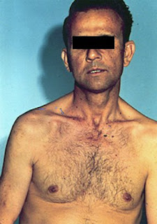
look at the eyes of this patient.
what is defferential diagnosis?

Friday, 6 June 2008

history taking


opening the interview:
introduce yourself appropriately and explain your role.
maintain attentive body position,good eye contact.

clinician:
"come in..."
"Hello,..Mr Osbourne, is it?" shakig hand with patient.
"common have a seat."
"nice to meet you."
"my name is Mr Steven, am a Urologist."

the main complaint:
obtain and record the main complaint in the patient own words.
let the patient finish the opening statment.
start with open-end questions.

clinician:
"I wonder if i can ask you a little about why are you here?,...and what problems you have been experiencing?"
"ok..,do you want to tell me more about that?"
"ok..,are there any other problems associated with that at all?"
"is there anytthing else you think of at the moment?"

strive for interchangeable responces, such as silence and noding.
use summerization and clarification.

clinician:
"so..am gathering obviously that the waterwork is getting worse and also the tiredness issue..."


close-ended questions provide details and they are useful in trying to build the case for certain diagnosis.
the "wh-" questions describe the attributes of the patient's symptoms and specify the story.

clinician:
"first of all,..HOW long these symptoms have been a problem for you?"
"WHEN was the first time you noticed that there is a problem at all?"

facilitation;
clinicain:
"..and then,..you saw your GP I think,is that right?"

summerize and recab,
clinican:
"can I actually ask about the main symptom again, you mentioned...."

shaw empathy:
clinician:
"I can imainge that can be very inconvenient at times.."

getting the details.
clinician:
"can i ask you when you go, is it painfull to pass urine at all?"
"..have you noticed blood in you urine..?"
"..do you got bony pain or joint pain?"

re summerize:
clininian:
"if you dont mind can I just summerize...."


history taking skills

Opening the Interview/Setting the Agenda

The Trouble with Physicians

In one study, physicians did not allow patients to complete their opening statements 69% of the time. The mean time until the first interruption was 18 seconds. Once interrupted, fewer than 2% of patients went on to complete their statements.

"Data are thus very much physician-determined, skewed toward problems that are biomedical in nature... It has been proposed that current interviewing practices are at odds with scientific requirements: They produce biased, incomplete data about the patient."

An Outline for the Opening the Interview

Setting the Stage

Goal: To establish a favorable context for the interview

  • Welcome the patient

  • Know and use the patient's name

  • Introduce and identify yourself

  • Ensure comfort and privacy

Chief Complaint/Setting the Agenda

Goal: To establish the agenda for the interview

  • Obtain list of all issues - avoid detail

    • Chief Complaint

    • Other complaints or symptoms

    • Specific requests (i.e. medication refills)

  • Patient's expectations for this visit

  • Ask the patient "Why now?"

Eliciting the Patient's Story

Goal: To establish a good flow of information

  • Open-ended questions initially

  • Encourage with silence, nonverbal cues, and verbal cues

  • Focus by paraphrasing and summarizing

Transition

Goal: To smoothly shift into physician-centered interviewing

  • Summarize interview up to that point

  • Verbalize your intention to make the transition

An Outline for the Rest of the Interview

Prioritize Problems

This should be clear from the transition summary.

Pursue the Most Urgent Problem (History of Present Illness)

  • Move from general to specific

  • Flow from open-ended to closed-ended questions

Pursue Other Problems as Time Permits

Review of Systems

Past Medical History

  • Allergies/Adverse Reactions

  • Medications/Immunizations

  • Major Medical or Psychiatric Problems/Major Surgeries

  • Last Menstrual Period/Pregnancies/Contraception (if female)

  • Smoking/Alcohol/Caffeine/Other Drugs

Other History (as appropriate)

  • Family/Social History

  • Occupational History

  • Sexual History


Parkinson’s disease


→Difficulty to start walking
→Quick shuffling gate with no arm swinging
→Difficulty to turn back

→Has expressionless face
→Low volume monotonus speech
→Has drooling
→There is titubation

→Pill rolling tremor
→Bradykinisia
→The arms showed lead-pipe rigidity at the elbow
Cog-weal rigidity at the wrist

→Gabellar tap is positive (Myerson’s sign)
→Signs are generally asymmetrical
→Hand writing show micrograghia

what comprise Parkinson's syndrome?
what are the causes of Parkinson's syndrome?
what do you know about Parkinson's plus syndromes?
talk about treatment and management.


NICE guidlines June 2006

exam question


look at this patient's face.
describe your findings.
what is your diagnosis?

if you picked the diagnoses,proceed as follow:
1-shake hands
2-inspect face carefully,note balding/wasting/ptosis/cataract
3-examin upper limbs/lower limbs:wasting and decrease reflexes
4-perform percusion myotonia
5-tell the examiner that you would like to examine for:gynaecomastia/testiculr atrophy.
6-ask the patient about dysphagia/recurrent chest infection/FHx
7-request investigations


internuclear ophthalmoplagia


click on the image to enlarge.

what are the causes of INO?
how would you investigate?

Carpal tunnel syndrome


how would you confirm the disgnosis?

foot drop


click on the image to enlarge.

foot drop is caused by weakness of the dorsoflexors of the foot and toes.

site and causes:
1-lumbosacral pluxus (pelvic malignancy)
2-sciatic nerve palsy (trauma,IM injection)
3-peroneal nerve palsy (what are the causes?)
4-neuomuscular junction(Myathenia Gravis )
5-muscles/myopathy(myotonic dystrophy)

what are the features of foot drop due to common peroneal nerve palsy?
1-wasting of the muscles in the lateral aspect of the leg
2-weakness of dorsoflexion +/- eversion
3-high seppage gait
4-loss of sensation in the lateral aspect of the leg and ddorsum of the foot.

eye examination

usefull powerpoint presentation about eye examination.

Thursday, 5 June 2008

examine this patient's legs


what is your diagnosis?
what is the mode of inheritance?

features:
wasting of the muscles of the calves and thigh that stops in the lower third of the thigh giving "inverted champagne bottle"appearance.
pus cavus
clawing of the toes
weakness of dorsiflexion
wasting and clawing of the muscles of the hand

look for:
lateral popliteal nerve thickening
enlarge greater auricular nerve
gate (high steppage gate due to foot drop)
ask about family history.

Stroke


there is right UMN weakness of the facial muscles.the right arm and leg are weak with increased tone and hyper-reflexia.the right planter is extensor.
he is dysphasic and there is sensory inattension.
this is right hemiplegia.

I would like to finish my examination by:
examin the visual field for homonymous hemianopia
check for caroted bruit
pulse for AF/heart for murmur
check the BP
dip the urine for suger

what are the causes of stroke?
what are the risk factors?

how would classify stroke?
Bamford classification 1991:
1-total anterior circulation stroke TACS
higher cerebral dysfunction +
homonymous visual field defect +
ipsilateral motor and/or sensory deficit
2-partial anterior circulation stroke PACS
2 out of 3 of TACS or higher cerebral dysfunction alone
3-lacunar stroke LACS
motor stroke or sensory stroke or sensory-motor stroke
4-posterior circulation stroke POCS
ipsilateral CN palsy with contralateral motor +/-sensory deficit or
bilateral motor+/-sensory deficit or
cerebellar dysfunction


can you list the features that are helpful t localize the site of hemiplagia?

examin this patient's eye


If you noticed ptosis, answer the following questions:
1-is ptosis complete or partial
2-is it unilateral or bilateral
3-is the pupil constricted ->Horner’s syndrome
Dilated->3rd nerve palsy
4-are extra ocular muscles involved?
5-is the eyeball sunken or not (enophthalmos)
6-is the light reflex intact (intact light reflex in Horner’s)

If the patient has Horner’s syndrome then proceed as follow:
Examine the supraclavicular area
Look for scar of cervical sympathectomy
Look for enlarge lymph nodes
Percuss for dullness (pancoast tumour)
Examine the neck
Carotid and aortic aneurysm
Tracheal deviation
Examine the hands
Small muscle wasting
Sensation
Clubbing
Ask about loss of sweating on one side of the face.
Look for evidence of brain stem vascular disease or demyelination

presentation:
this patient has miosis,enophthalmos and left ptosis.
this is left Horner's syndrome.

what is Horner's syndrome?
Horner syndrome results from an interruption of the sympathetic nerve supply to the eye, and is characterized by the classic triad of miosis (ie, constricted pupil), partial ptosis, and loss of hemifacial sweating (ie, anhidrosis).
Horner syndrome isn't a disease but a sign of an underlying — often serious — disorder

what are the causes of Horner's syndrome?

wasting of the small muscles of the hand


click image to enlarge.
examin this patient's hand.
describe your findings.
what are the causes of unilateral/bilateral wasting?

there is bilateral wasting and weakness of the thenar and hypothenar in addition to dorsal guttering.there is no fasciculation.no evidance of Horner's syndrome.the patient is unable to unbutton his clothes.


Wednesday, 4 June 2008

exam question 7


describe.
what is your diagnosis?
what other signs you would look for?

renal transplantation

exam question:
patient with renal transplant and arteriovenous fistula,did not had palpable kidneys.
what are the causes of renal failure?
discussion about polycystic kidney disease(presentation/complications)

Polycystic kidney disease:
AD chromosome 16
Prevalence 1:1000
The third most common cause of CRF in the UK

see presentation below.
what are the most used immunosuppresive medications and its saide effects?

typical case


click on the image to enlarge


look out for this in the abdomin station.
if you found it,you should score 4!

renal...

hepatosplenomegaly


exam question:
hepatosplenomegaly,signs of chronic liver disease.
asked to discuss causes of portal hypertension.
asked about the management of a patient with acute hepatic encephalopathy.

causes of hepatosplenomegaly:
1-Infection:
Acute viral hepatitis
Infectious mononucleosis
Cytomegalovirus
2-Haematological disease:
Myeloproliferative disorders :CML,Myelofibrosis.
Lymphoproliferative disorders:CLL, Hodgkin's Lymphoma
Anaemia:Pernicious anaemia ,Sickle cell anaemia,Thalassaemia
3-Chronic liver disease and portal hypertension:
Chronic active hepatitis
4-Amyloidosis
5-Acromegaly
6-Systemic lupus erythematosus

another exam question:
anaemic patient with hepatosplenomegaly.
most likely diagnosis:myelfibrosis
discuss.

Tuesday, 3 June 2008

splenomegaly


exam question:
young black man with massive splenomegaly.no lymph nodes.asked for the single most likely diagnosis.

causes for splenomegaly:
1-Neoplastic
Lymphoproliferative: CCL, lymphoma
Myeloproliferative: CML, myelofibrosis
2-Inflammatory
Amyloidosis SLE
Sarcoidosis RA
3-Infective
Glandular fever
Hepatitis
Chronic malria/Brucelosis
SBE
4-Others
PRV cirrhosis
ITP storage disease

in which conditions the spleen is massively enlarged?

another exam question:
large isolated splenomegaly,no anaemia,no lymphadenopathy.
what is the most likely diagnosis?
how would you investigate?

examination tip

ascites


exam question:
middle age male unwell with nausea and vomiting and abdominal distension,juandiced,ecchymoses,hepatosplenomegaly with ascites.had a urinary catheter.
asked to give various defferentials.
asked about investigation and management of ascites.

ascites is the pathological presence of fluid within the peritoneal cavity.
common causes:
1-liver cirrhosis with portal hypertension
2-malignacy
3-congestive cardiac failure

investigatons:
ascetic tab and send fluid for:
biochemstry
cytology
cell count,culture and gm stain

use the protein level to defferentiate between exudate and transudate.
exudate has protein content of 25g/L
serum-ascites albumin gradient correlate directly with the portal press.
SAAG gradient>11g/L indicate portal hypertension with transudate
SAAG gradeint<11g/L indicate exudate

lines of management:
1-bed rest ?of little value
2-soduim restriction
3-water restriction
4-diuretcs therapy
5-paracentesis
6-shunt

what are the complications of ascites?
what is the pathophysiology of ascites?

Asthma

if you got a patient with asthma,you are likey to be asked about the stepwise treatment:
Step1
Short acting beta agonist as required
e.g salbutamoul PRN

Step 2
Short acting beta agonist as required
+ Standard dose inhaled corticosteroids
e.g salbutamoul inh PRN
Beclomethasone inh regular

Step 3
Short acting beta agonist as required
+standard dose inhaled corticosteroids regular
+long acting beta agonist regular
e.g salbutamoul inh PRN
Beclomethasone inh regular
Salmetrol inhalers regular

If no response, consider increase the dose of regular inhaled corticosteroids to the upper limit of standard dose.

Step 4
Short acting beta agonist as required
+standard dose inhaled corticosteroids regular
+long acting beta agonist regular
+6 weeks trial of one or more of:
Leukotrienes receptors antagonists
Modified released thiophylline
Modified released oral beta agonist

Step 5
Add prednisolone tab

what are the features of acute svere asthma and how would you manage that?
what are the indications for mechanical ventilation?

Monday, 2 June 2008

scar...again


what is the name of this scar?
what are the indication for liver transplant?
what is the most likely aetiology in this patient?

Any patient with documented fulminant hepatic failure, decompensated cirrhosis, or hepatocellular carcinoma within defined criteria (no single lesion greater than 5 cm or no more than three lesions, the largest ≤3 cm) is a potential candidate for liver transplantation.
Any patient with one of the defined complications of end-stage liver disease (eg ascites, variceal bleeding, encephalopathy or hepatocellular carcinoma) and/or a MELD score of 10 should be considered for referral to a transplant center.


BSG guidelines for liver transplant, click here

the PACES liver

Addison's disease


there is generalized pegmentation,most marked in scars,skin crease and buccal mucosa.

examin the abdomen for adrenal scar(Nelson syndrome)
check the BP lying and standing

how would you confirm the diagnosis?
Random cortisol
Short synacthin test (normal cortisol rise above 600 nmol/l or increase by 300 nmol/l)
ACTH level or long synacthin test

what is the most common cause?
mention other caues of hyperpegmentaion.
what is Schmidt's syndrome?

Cushing's syndrome


this patient has a moon face with acne,there is truncal obesity and buffalo hump.the skin is thin with excessive bruising and there is purple striae in the andomen.there is proximal muscle weakness.

causes of Cushing syndrome:
Sustained over production of cortisol

Cushing syndrome (non-ACTH dependent)
1-Adrenal adenoma
2-Adrenal carcinoma
3-Glucocorticoid administration

Cortisol ^ , ACTH v

Cushing disease (ACTH dependent)
1-Pit dependent
2-Ectopic ACTH producing tumour
3-ACTH administration

Cortisol ^ , ACTH ^

NB, pigmentation occur with ACTH dependent causes

Features:
1)central obesity
2)hirsutism
3)recurrent infections
4)osteoporosis ( can cause vertebral collapse)
5)olegomenorrhoea
6)hypokalaemia (due to mineralocoticoid activity )
7)water retension ( ADH )
8)hypoglycaemia ( -ve to insulin )
9)hypertension
10)proximal myopathy and wasting

Investigations:

1-screening:
24 hrs free urinary cortisol
Mid night dexamethasone suppression test

2-cofirmation test
For +ve screening test
Depend on demonstration of inappropriate cortisol secretion not suppressed by exogenous glucocorticoid.
Normal individual suppress cortisol to less than 50 nmole/L
low/high dose dexamethasone suppression test

discuss the treatment/managment of this patient.

osteoarthritis


examine this patient's hand.
desribe your findings.

there is Heberden's nodules at the base of DIJ and Buuchard's nodules at the PIJ.
there squiring of the first metacapal due to subluxation.
now assess functional status.
examin for hip and knee involvement.

what are the typical radiological features?
how would you manage this patient?

Psoriatic arthropathy


asymetrical arthropathy involving mainly the DIJ.
there is pitting of the nail and onycholysis.
(look for patches of psoiasis in the skin)

radiological features:
1-fluffy periostitis
2-destruction of the small joints
3-pencil cup appearance
4-deformity

what are the different types of psoriaric arthropathy?
what are the lines of management?

Gout


asymetrical swelling affecting the small joints of the hand (and feet),the joints are severely deformed.there are tophi formation in the halex of the ear.

its a metabolic disorder of purine metabolism,characterized by hyperuricaemia due to either overproduction or under excretion of uric acid.

main risk factors:
age
diabetes
high BMI (obesity)
high protein diet
high alcohol consuption
hypertension
hyperlipidaemia
family Hx
drug Hx

what the main clinical manifestations?
what is the defferential diagnosis?
how would you investigate this patient?
what are the lines of management?

exam question 6


describe.
what is your diagnosis?
what other skin lesions associated with this condition?
what is the mode of inheretance?

exam question 5


describe.
what is your diagnosis?
what questions would you like to ask?
read more about this condition

exam question 4


describe your findings.
what is your diagnosis?
how would you investigate?
how would you treat this patient?

read more...

should score 4 in psoriasis


describe your findings.
there are patches of psoriasis over the trunk of the body and the bony promenance.
the plaques are circular ,well defined adge with salivary scales.
there is no asymetrical arthropathy present.

what are the types of psoiasis?
1-plaque
2-guttate
3-pustular
4-erythrodermal

what factors can exacrebate psoriasis?
1-drugs e.g B blockers,ACE inhibitors,lithuim,antimalarials and alcohol
2-stress
3-infections
4-trauma to the skin
5-sun light

what are the lines of management?
no cure,only supportive treatment.
indication for treatment:
1-local symptoms
2-cosmoitc problems
3-both
goal of treatment:
to decrease the extent and severity of the condition to the point that it no longer interfer substantially with patient occupation,personal or social life.

therapy:
1-topical treatment
2-systemic treatment (phototherapy or systemic drugs)
read more

Sunday, 1 June 2008

scleroderma


the skin over the fingers is smooth, shiny and tight
there is digital ischemia and ulceration with pulp infarction and atrophy
there is dilated nail fold capillaries with nail fold infarct and ragged cuticle
there is calcinosis.
now comment on any features on the face.
what are the other organs involved?
what are the varient of scleroderma?
what are the lines of manahement?


read more.